The picture of an x ray machine comes equipped with advanced digital detectors that transform X-ray energy into high definition images of incredible detail. The system's design makes it easy to use and facilitates quick image capturing. The picture of an x ray machine system can be connected effortlessly to hospital information systems that enable the secure transfer of data. The system's robust design provides support for long-term use within healthcare settings.

The picture of an x ray machine is commonly used in medical imaging to examine skeletal trauma, lung disease, and dental anatomy. The picture of an x ray machine assists physicians in diagnosis of fractures, infection, and degenerative disease. The picture of an x ray machine is also used in orthopedic surgery intraoperatively. In emergency medicine, it provides rapid diagnostic information that allows clinicians to assess trauma and internal injury rapidly.
Future versions of the picture of an x ray machine will combine energy-efficient technology with high-resolution imaging. Predictive analytics integration will enable early disease detection and personalized screening. Global telemedicine networks will also be enabled by the picture of an x ray machine, extending access to diagnosis in underserved populations.
Regular upkeep of the picture of an x ray machine enhances operating efficiency and patient safety. Regular exposure level checks and image acuteness tests guarantee reproducible output. The picture of an x ray machine should be operated by well-trained operators who observe cleaning and handling protocols to reduce wear and prolong service life.
The picture of an x ray machine represents an important diagnostic tool that functions by using controlled X-ray radiation to create images of the bones, organs, and internal structures of the body. The equipment assists healthcare providers in diagnosing ailments such as fractures and infections with high accuracy. The picture of an x ray machine equipment is usually found in hospitals and dental clinics as it provides efficient imagery services that aid in comprehensive diagnoses. The equipment's efficiency makes it an important aspect of modern medical facilities.
Q: What makes an x-ray machine different from a CT scanner? A: An x-ray machine captures a single 2D image, while a CT scanner takes multiple x-rays from different angles to create 3D cross-sectional views. Q: How is image quality measured in an x-ray machine? A: Image quality depends on factors like contrast, resolution, and exposure settings, which are adjusted based on the target area being examined. Q: What power supply does an x-ray machine require? A: Most x-ray machines operate on high-voltage power systems, typically between 40 to 150 kilovolts, depending on their intended use. Q: Can x-ray machines be used for dental imaging? A: Yes, specialized dental x-ray machines provide detailed images of teeth, jaws, and surrounding structures to support oral health assessments. Q: How does digital imaging improve x-ray efficiency? A: Digital systems allow instant image preview, faster diagnosis, and reduced need for retakes, improving workflow efficiency in clinical environments.
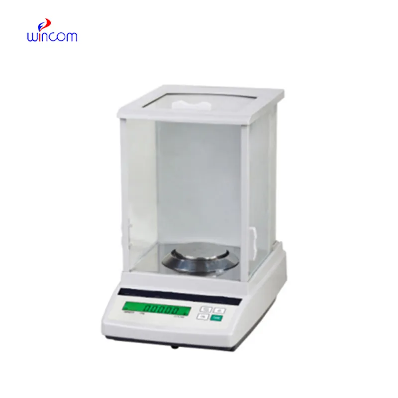
The microscope delivers incredibly sharp images and precise focusing. It’s perfect for both professional lab work and educational use.
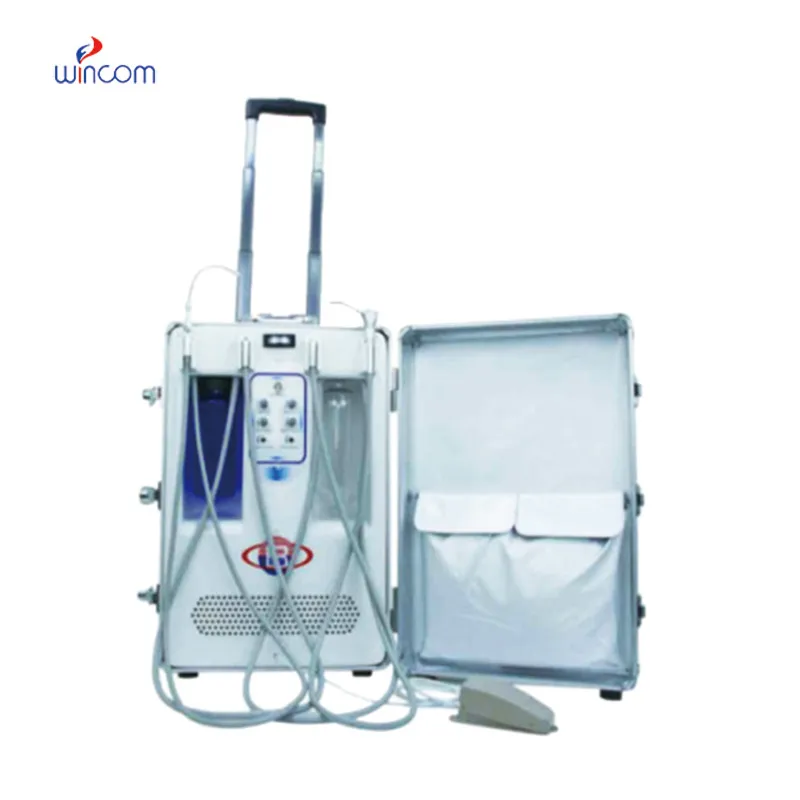
The delivery bed is well-designed and reliable. Our staff finds it simple to operate, and patients feel comfortable using it.
To protect the privacy of our buyers, only public service email domains like Gmail, Yahoo, and MSN will be displayed. Additionally, only a limited portion of the inquiry content will be shown.
We’re currently sourcing an ultrasound scanner for hospital use. Please send product specification...
I’m looking to purchase several microscopes for a research lab. Please let me know the price list ...
E-mail: [email protected]
Tel: +86-731-84176622
+86-731-84136655
Address: Rm.1507,Xinsancheng Plaza. No.58, Renmin Road(E),Changsha,Hunan,China

 af
af
 es
es
 ar
ar
 tr
tr
 sw
sw
 pt
pt
 th
th
 ur
ur
 bn
bn
 ne
ne
 vi
vi
 km
km
 lo
lo
 de
de
 ru
ru
 fi
fi
 nl
nl
 fa
fa
 fr
fr
 ko
ko