Through a combination of novel imaging software and the ultrasound machine ge, the professionals can be very precise with the details of the anatomy they have captured and even more so with the accuracy of the final result. An ergonomic design of the device contributes to the reduction of operator fatigue during long use. The device, the ultrasound machine ge, also comes with the ability to connect to more than one probe thus giving it the flexibility required when catering to various diagnostic needs. Besides that, the data export and connection functions of the device help to decrease the time taken in report-generating which is done through image sharing.
The ultrasound machine ge is the workhorse in oncology because it assists in the accurate locating of tumors and keeping an eye on the treatment progress. It helps in making the right calls concerning benign versus malignant lesions in breast and thyroid cases. The ultrasound machine ge is also there in support of interventional procedures such as guided aspirations and injections.
The future of the ultrasound machine ge may include integration with artificial intelligence for automatic analysis of images. Increased portable functionality and wireless communications may improve remote accessibility in healthcare. The ultrasound machine ge may include deeper imaging capabilities and rapid data transfer with cloud storage that fosters collaboration on a global medical scale.
The ultrasound machine ge needs to be maintained properly to ensure it always provides high-quality images. The probes should never be dropped or immersed in liquid against the recommended standards. Care should be taken when handling the control board to avoid mechanical wear and tear. Updation of the firmware and testing of the ultrasound machine ge ensure smooth functionality during practical operations.
With advanced transducer technology, the ultrasound machine ge delivers reliable and high-resolution imaging capability. It allows healthcare providers to see anatomical structures with unmatched clarity and speed. The ultrasound machine ge enhances workflow efficiency in hospitals, clinics, and mobile medical facilities. With rugged construction and digital connectivity, it makes integration into existing healthcare systems easy.
Q: What is the primary function of an ultrasound scannert? A: Ultrasound scanners are designed to create real-time images of internal organs, tissues, and blood flow using high-frequency sound waves. Q: How does the ultrasound scannert ensure clear imaging results? A:It uses advanced converter technology and digital processing to enhance image clarity and contrast. Q: In what medical fields is the ultrasound scannert commonly used? A: It is widely used in obstetrics, cardiology, urology, radiology, and emergency medicine. Q: Is the ultrasound scannert safe for repeated use? A: Yes, it is non-invasive and does not emit radiation, making it safe for frequent diagnostic applications. Q: Can the ultrasound scannert store and share imaging data? A: Yes, it supports data storage, retrieval, and digital transfer for easy integration with hospital systems.
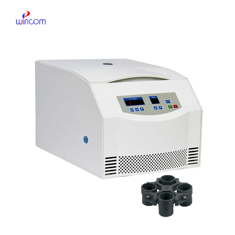
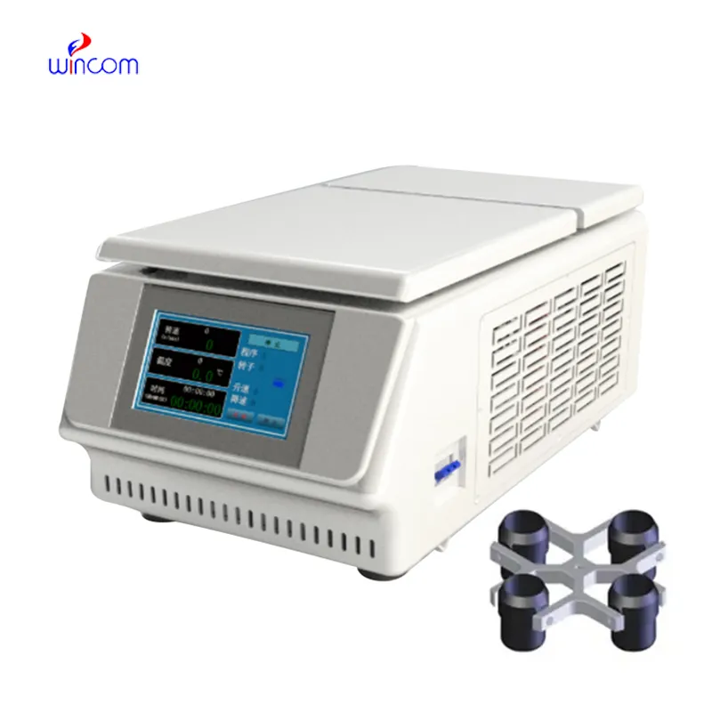
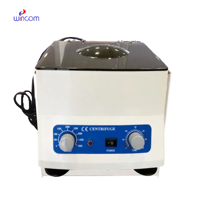
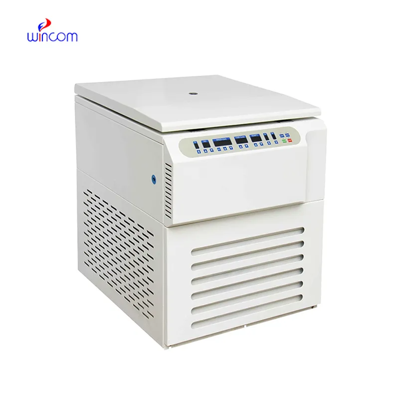
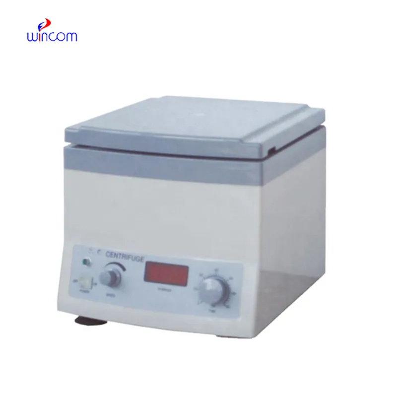
We’ve used this centrifuge for several months now, and it has performed consistently well. The speed control and balance are excellent.
I’ve used several microscopes before, but this one stands out for its sturdy design and smooth magnification control.
To protect the privacy of our buyers, only public service email domains like Gmail, Yahoo, and MSN will be displayed. Additionally, only a limited portion of the inquiry content will be shown.
We’re interested in your delivery bed for our maternity department. Please send detailed specifica...
We are planning to upgrade our imaging department and would like more information on your mri machin...
E-mail: [email protected]
Tel: +86-731-84176622
+86-731-84136655
Address: Rm.1507,Xinsancheng Plaza. No.58, Renmin Road(E),Changsha,Hunan,China

 af
af
 es
es
 ar
ar
 tr
tr
 sw
sw
 pt
pt
 th
th
 ur
ur
 bn
bn
 ne
ne
 vi
vi
 km
km
 lo
lo
 de
de
 ru
ru
 fi
fi
 nl
nl
 fa
fa
 fr
fr
 ko
ko