rp-hplc enables labs to separate and analyze intricate mixtures with utmost precision. Through a seamless connection with current detectors, the method provides detailed profiling of both chemical and biological substances. The researchers and therapists trust rp-hplc for the purposes of monitoring outcomes of experiments, method development, and cross-analyses accuracy. Its strength in dealing with various kinds of samples renders it an indispensable device in both the research and the clinical settings, thus improving reproducibility and backing up the struggling with more complex scientific and medical inquiries.

In rp-hplc used to analyze metabolic profiles and biomarkers during clinical research laboratories. It enables the identification of disease markers and monitoring of biochemical changes over time through the separation of small molecules and proteins. rp-hplc also facilitates the study of drug absorption and distribution, toxicity testing, and hospital-based clinical trials and thus making it possible to monitor patient responses to therapies in great detail while at the same time ensuring the accuracy and reliability of the analytical results.
The future of rp-hplc stresses the integration of hospital information systems and electronic medical records. The analysis of patient samples will be automatically included in the clinical workflows. Increased automation, AI-based interpretation, and better sensitivity will put rp-hplc at the center of the laboratory operations and patient care that is focused on the patient's needs.
The hospital labs keep their rp-hplc by adopting diligent handling and preventive maintenance. The regular examination of the columns, pumps, and connectors, along with the correct use of the solvents, aids in eliminating the problems of blockages and pressure. The lab staff is recommended to observe the cleaning and calibration according to the manufacturer's manual. The, such practices are applied, they bring about the benefits of long-term reliability, consistent separation quality, and accurate analytical outcomes in both clinical and experimental workflows.
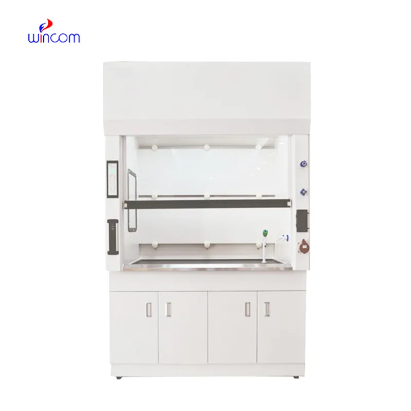
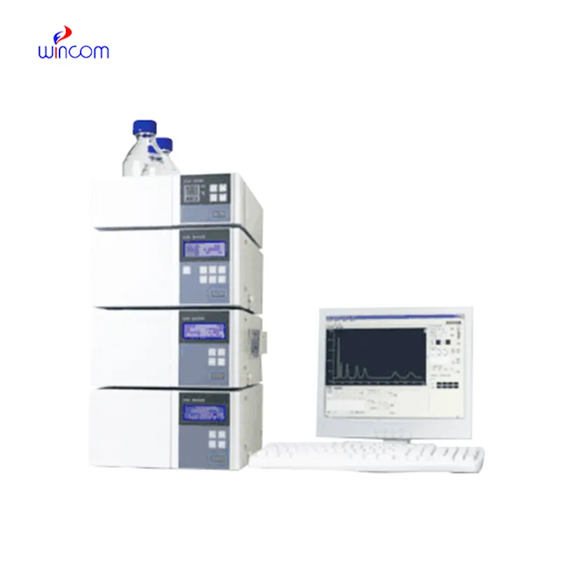
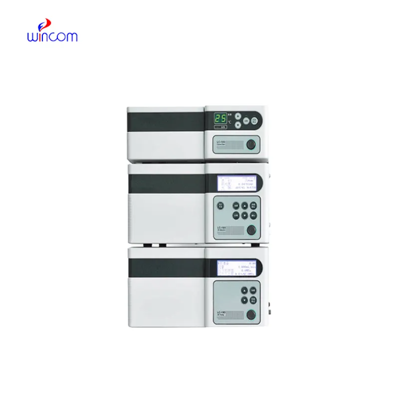
rp-hplc is commonly employed in laboratories to separate, identify, and quantify chemical compounds. The sample mixture is put through the columns along with the stationary phases and the different components interact with the stationary phase, thus the separation is done accurately. This process not only gives high resolution but also reproducibility thus it is a must-have tool for the research works in the area of drugs, pollution, and food control. Subsequently, when coupled with sensitive detectors, rp-hplc facilitates the precise measurement of minor concentrations. The method versatility produces so much that it has become a necessity in a routine analysis and complex research applications where it is positioned as an essential instrument in contemporary analytical chemistry and experimental workflows.
Q: Do you need special training for HPLC operation? A: The answer is yes, training is a prerequisite to accurately and safely using pumps, columns, and detectors. Q: What type of maintenance does HPLC have? A: It requires cleaning, flushing, and inspection of all components as well as calibrating. Q: Is it possible to use HPLC in drug monitoring? A: Sure, it is a common practice in hospitals to monitor the levels of therapeutic drugs and also to identify metabolites in the samples taken from the patients. Q: What is the duration of analysis using HPLC in a typical case? A: The analysis time can range from a few minutes to more than an hour depending on the nature of the sample and the kind of column used. Q: Is HPLC a good choice for environmental testing? A: Yes, it can be used to find out the presence of pollutants, pesticides, and other harmful substances in water, soil, and air samples.
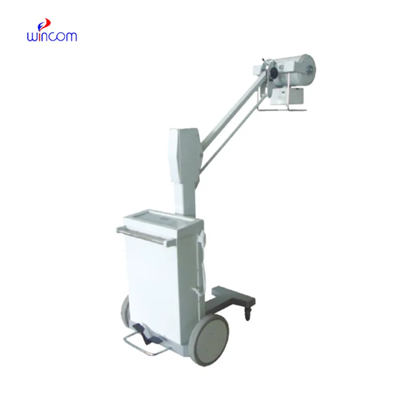
This x-ray machine is reliable and easy to operate. Our technicians appreciate how quickly it processes scans, saving valuable time during busy patient hours.
We’ve been using this mri machine for several months, and the image clarity is excellent. It’s reliable and easy for our team to operate.
To protect the privacy of our buyers, only public service email domains like Gmail, Yahoo, and MSN will be displayed. Additionally, only a limited portion of the inquiry content will be shown.
We are planning to upgrade our imaging department and would like more information on your mri machin...
Could you share the specifications and price for your hospital bed models? We’re looking for adjus...
E-mail: [email protected]
Tel: +86-731-84176622
+86-731-84136655
Address: Rm.1507,Xinsancheng Plaza. No.58, Renmin Road(E),Changsha,Hunan,China

 af
af
 es
es
 ar
ar
 tr
tr
 sw
sw
 pt
pt
 th
th
 ur
ur
 bn
bn
 ne
ne
 vi
vi
 km
km
 lo
lo
 de
de
 ru
ru
 fi
fi
 nl
nl
 fa
fa
 fr
fr
 ko
ko