Through a combination of novel imaging software and the pink fetal doppler, the professionals can be very precise with the details of the anatomy they have captured and even more so with the accuracy of the final result. An ergonomic design of the device contributes to the reduction of operator fatigue during long use. The device, the pink fetal doppler, also comes with the ability to connect to more than one probe thus giving it the flexibility required when catering to various diagnostic needs. Besides that, the data export and connection functions of the device help to decrease the time taken in report-generating which is done through image sharing.

The vast clinical applications of the pink fetal doppler technology made it possible for nephrology to monitor kidney function efficiently and detect abnormalities in kidney structure. In the frontiers of endocrinology, the obtained data can reveal even the smallest nodules in the glands. The pink fetal doppler is also a surgical device for blood flow patterns and vessel integrity.

Through continued innovations in digital technology, the pink fetal doppler can be expected to improve and extend its applications within preventive medicine and telemedicine. The next generation of such technologies will facilitate collaboration among experts in real-time using cloud-imaging solutions. The pink fetal doppler can also work within wearables that include biosensors.

In order to retain the accuracy of the pink fetal doppler, it is important for operators to check the cables and connections of the transducers for evidence of wear. After each use, the surfaces should be wiped clean using non-abrasive cleaners. The pink fetal doppler should be turned off properly and covered to prevent dust from collecting. Regular checks by trained personnel should be done.
Used in hospitals and clinics, the pink fetal doppler provides immediate visual feedback for a variety of medical evaluation uses. Converting sound waves into live images, the pink fetal doppler allows physicians to easily detect abnormalities. The pink fetal doppler assists with making diagnostic processes safer in addition to improving patient outcomes. It possesses an ergonomic shape alongside digital integration capabilities that support simple data sharing and medical record documentation.
Q: What imaging modes are available on the ultrasound scannert? A: It supports multiple modes such as B-mode, M-mode, and color Doppler for diverse diagnostic applications. Q: How does the ultrasound scannert improve diagnostic accuracy? A: By providing high-resolution images and real-time feedback, it enables more precise medical evaluations. Q: Can the ultrasound scannert be used in field or remote settings? A: Yes, its portable versions are designed for mobility and can be used in clinics, hospitals, or mobile healthcare units. Q: What kind of display does the ultrasound scannert use? A: It typically features a high-definition digital display that enhances image visualization and readability. Q: How is data from the ultrasound scannert managed? A: The device allows secure storage, easy access, and export of imaging data through USB or network connections.
We’ve been using this mri machine for several months, and the image clarity is excellent. It’s reliable and easy for our team to operate.
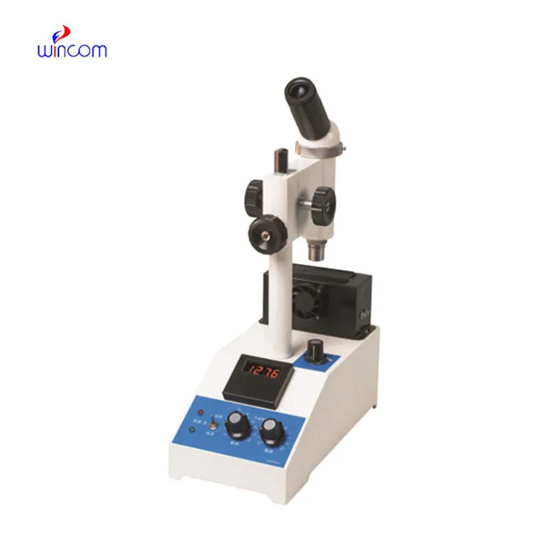
The microscope delivers incredibly sharp images and precise focusing. It’s perfect for both professional lab work and educational use.
To protect the privacy of our buyers, only public service email domains like Gmail, Yahoo, and MSN will be displayed. Additionally, only a limited portion of the inquiry content will be shown.
Could you please provide more information about your microscope range? I’d like to know the magnif...
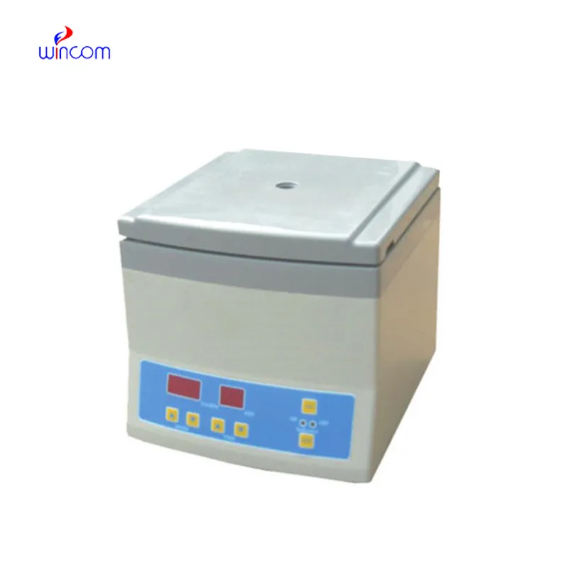
Hello, I’m interested in your centrifuge models for laboratory use. Could you please send me more ...
E-mail: [email protected]
Tel: +86-731-84176622
+86-731-84136655
Address: Rm.1507,Xinsancheng Plaza. No.58, Renmin Road(E),Changsha,Hunan,China

 af
af
 es
es
 ar
ar
 tr
tr
 sw
sw
 pt
pt
 th
th
 ur
ur
 bn
bn
 ne
ne
 vi
vi
 km
km
 lo
lo
 de
de
 ru
ru
 fi
fi
 nl
nl
 fa
fa
 fr
fr
 ko
ko