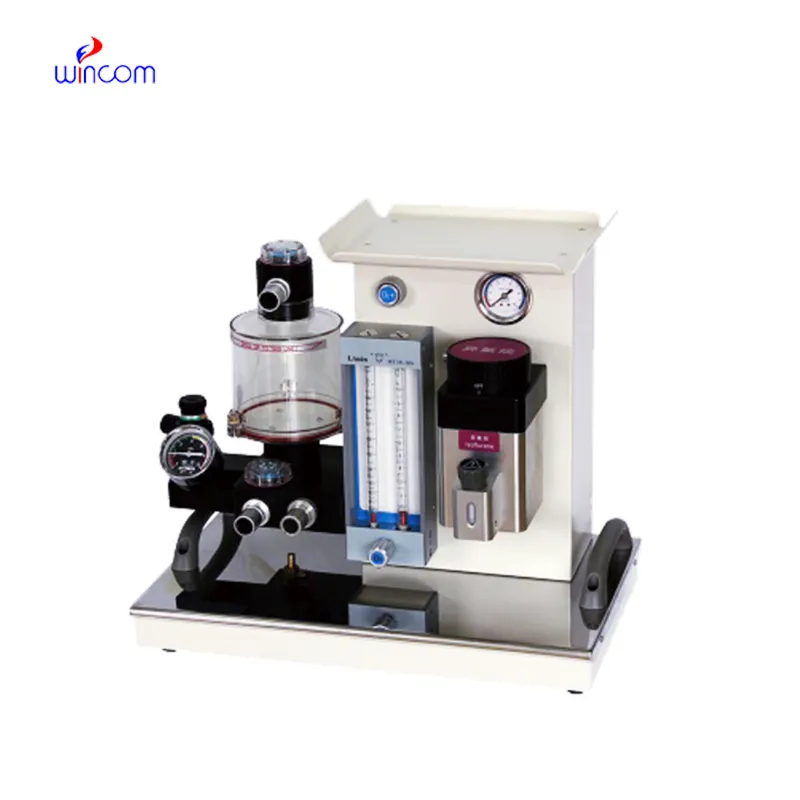
mri anesthesia machine assists hospitals in providing exact anesthesia by constantly observing patient respiration and oxygenation. The integrated system of mri anesthesia machine is made up of vaporizers, flow meters, and alarms that work together to control the level of anesthetic and ensure safety for the patient. Anesthesiologists in operating rooms and intensive care units rely on this device to perform real-time monitoring of airway pressures and ventilation parameters. Manifold and very precise gas delivery and respiratory monitoring are the requirements for laboratory-based research studies, where mri anesthesia machine already has its application. The combination of safety features and continuous physiological feedback provides consistent and reliable anesthesia management in both clinical and experimental settings.

mri anesthesia machine finds its place in diagnostic and interventional procedures where sedation is needed but not full surgical anesthesia. Imaging-guided interventions or endoscopic examinations usually require controlled sedation and also airway support. The equipment gives the practitioners the power to control the patient's respiration, at the same time keeping the anesthetic at the correct level. Its usage provides relief for the patient and also enhances the precision of the procedure. By enabling non-surgical medical methods, mri anesthesia machine increases the area of anesthesia management from the operating room to different hospital wards.
In the future, mri anesthesia machine will very probably add the features of ventilation that are more automated and suited to patients' breathing habits. Smart algorithms could help the doctors by changing the settings of ventilation according to the constant feedback. This development could lessen the need for human control in long operations without losing the accuracy of respiratory control. The same consistency in anesthesia delivery could be achieved in hospitals and laboratories doing research. These advancements are characteristic of the new medical environments where the anesthesia equipment will be more adaptive and responsive.
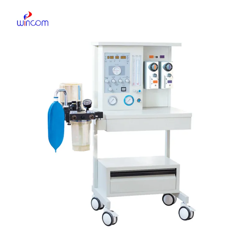
The proper care of mri anesthesia machine starts with weekly inspection before and after daily clinical use. The gas pipelines, connectors, and flow meters should be checked by the hospital staff to make sure they are working correctly. Cleaning and replacing of breathing circuits and masks should be done according to infection control protocols. Regular calibration of monitoring components is a way to always have accurate readings. In high-traffic operating rooms, stable maintenance schedules cut down on sudden breaks. Hospitals can prolong the lifespan of mri anesthesia machine and keep anesthesia delivery reliable by using inspection schedules that are structured.
In pediatric and neonatal care, mri anesthesia machine is the equipment that accurately provides the anesthesia adjusted to the small patients. The device's monitoring systems follow the most sensitive patterns of respiration and oxygen level, which give anesthesiologists the opportunity to do precise alterations. The safe and controlled administration of anesthetic gases is very important in these specialized hospital departments. By offering trustworthy monitoring and ventilation, mri anesthesia machine keeps the patient stable during surgical procedures.
Q: What gases does the Anesthesia Machine make use of? A: The regular gases are oxygen, nitrous oxide, and volatile anesthetic agents. Q: Is the Anesthesia Machine suitable for long surgeries? A: Yes, it was designed to maintain stable anesthesia throughout long operations. Q: Under machine control how does the anesthesia concentration occur? A: The vaporizers dispense a precisely-measured amount of anesthetic gas to the patient. Q: Could the machine be used for pediatric patients? A: Yes, but only if the settings are modified by the experienced medical personnel. Q: Is there a built-in alarm system for the machine? A: Yes, alarms are present to alert the staff to changes in pressure, gas flow, or oxygen levels.
We’ve used this centrifuge for several months now, and it has performed consistently well. The speed control and balance are excellent.
This ultrasound scanner has truly improved our workflow. The image resolution and portability make it a great addition to our clinic.
To protect the privacy of our buyers, only public service email domains like Gmail, Yahoo, and MSN will be displayed. Additionally, only a limited portion of the inquiry content will be shown.
I’d like to inquire about your x-ray machine models. Could you provide the technical datasheet, wa...
I’m looking to purchase several microscopes for a research lab. Please let me know the price list ...
E-mail: [email protected]
Tel: +86-731-84176622
+86-731-84136655
Address: Rm.1507,Xinsancheng Plaza. No.58, Renmin Road(E),Changsha,Hunan,China

 af
af
 es
es
 ar
ar
 tr
tr
 sw
sw
 pt
pt
 th
th
 ur
ur
 bn
bn
 ne
ne
 vi
vi
 km
km
 lo
lo
 de
de
 ru
ru
 fi
fi
 nl
nl
 fa
fa
 fr
fr
 ko
ko