The fetal heart doppler combines state-of-the-art digital imaging algorithms that boost contrast and depth perception, which leads to soft tissues being visualized more clearly. The intelligent interface features customizable scanning modes and user profiles for easier operation. Due to its low power consumption and sturdy construction, the fetal heart doppler maintains performance that is even in high-demand clinical environments.

The fetal heart doppler has become a necessity for internal medicine as it provides real-time imaging during fluid drainage and biopsy guidance. It finds a place in critical care too for instant bedside evaluations. The fetal heart doppler is also utilized by veterinary surgeons to monitor the health of a patient animal, thus proving its utility beyond human medicine.

The fetal heart doppler can look forward to getting advantages from miniaturization and wearable technologies. Portable or handheld versions of the fetal heart doppler will become more widespread to facilitate quick diagnoses in rural as well as emergency setups. The integration of telemedicine services will thus facilitate concurrent consultations via the fetal heart doppler.

The fetal heart doppler require a certain set of procedures when it comes to handling them. The images on the display should always be cleaned with soft, lint-free cloths. The probe membranes should always be checked for the absence of cracks. The fetal heart doppler also require calibration verification.
With advanced transducer technology, the fetal heart doppler delivers reliable and high-resolution imaging capability. It allows healthcare providers to see anatomical structures with unmatched clarity and speed. The fetal heart doppler enhances workflow efficiency in hospitals, clinics, and mobile medical facilities. With rugged construction and digital connectivity, it makes integration into existing healthcare systems easy.
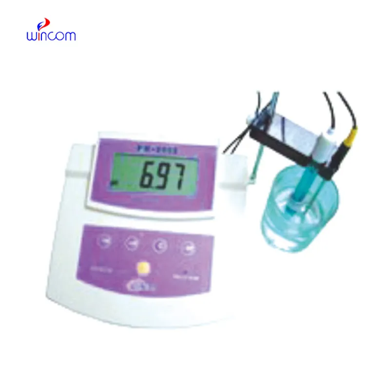
Q: What imaging modes are available on the ultrasound scannert? A: It supports multiple modes such as B-mode, M-mode, and color Doppler for diverse diagnostic applications. Q: How does the ultrasound scannert improve diagnostic accuracy? A: By providing high-resolution images and real-time feedback, it enables more precise medical evaluations. Q: Can the ultrasound scannert be used in field or remote settings? A: Yes, its portable versions are designed for mobility and can be used in clinics, hospitals, or mobile healthcare units. Q: What kind of display does the ultrasound scannert use? A: It typically features a high-definition digital display that enhances image visualization and readability. Q: How is data from the ultrasound scannert managed? A: The device allows secure storage, easy access, and export of imaging data through USB or network connections.
The microscope delivers incredibly sharp images and precise focusing. It’s perfect for both professional lab work and educational use.
The water bath performs consistently and maintains a stable temperature even during long experiments. It’s reliable and easy to operate.
To protect the privacy of our buyers, only public service email domains like Gmail, Yahoo, and MSN will be displayed. Additionally, only a limited portion of the inquiry content will be shown.
We’re currently sourcing an ultrasound scanner for hospital use. Please send product specification...
I’d like to inquire about your x-ray machine models. Could you provide the technical datasheet, wa...
E-mail: [email protected]
Tel: +86-731-84176622
+86-731-84136655
Address: Rm.1507,Xinsancheng Plaza. No.58, Renmin Road(E),Changsha,Hunan,China

 af
af
 es
es
 ar
ar
 tr
tr
 sw
sw
 pt
pt
 th
th
 ur
ur
 bn
bn
 ne
ne
 vi
vi
 km
km
 lo
lo
 de
de
 ru
ru
 fi
fi
 nl
nl
 fa
fa
 fr
fr
 ko
ko