The fetal doppler at 13 weeks combines state-of-the-art digital imaging algorithms that boost contrast and depth perception, which leads to soft tissues being visualized more clearly. The intelligent interface features customizable scanning modes and user profiles for easier operation. Due to its low power consumption and sturdy construction, the fetal doppler at 13 weeks maintains performance that is even in high-demand clinical environments.

The vast clinical applications of the fetal doppler at 13 weeks technology made it possible for nephrology to monitor kidney function efficiently and detect abnormalities in kidney structure. In the frontiers of endocrinology, the obtained data can reveal even the smallest nodules in the glands. The fetal doppler at 13 weeks is also a surgical device for blood flow patterns and vessel integrity.

The fetal doppler at 13 weeks should integrate with intelligent diagnostic ecosystems and communicate effortlessly with smartphones and electronic records. The synchronized exchange of data in real-time should enable constant patient observation. The next version should focus on improved design, better processing power of artificial intelligence algorithms, and enhanced reconstruction functions.

In order to retain the accuracy of the fetal doppler at 13 weeks, it is important for operators to check the cables and connections of the transducers for evidence of wear. After each use, the surfaces should be wiped clean using non-abrasive cleaners. The fetal doppler at 13 weeks should be turned off properly and covered to prevent dust from collecting. Regular checks by trained personnel should be done.
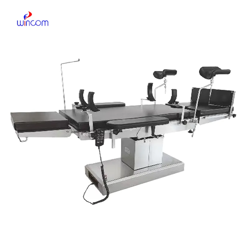
The fetal doppler at 13 weeks is more accurate in diagnostics as it captures high-resolution images of organs, tissues, and blood vessels. Design-wise flexible, it is used extensively in obstetrics, cardiology, urology, and musculoskeletal tests. Its portability and simplicity enable medical practitioners to make quick and precise evaluations. The fetal doppler at 13 weeks makes work processes more efficient and allows for the delivery of superior patient care through real-time visualization.
Q: What are the main maintenance requirements for the ultrasound scannert? A: Regular cleaning, proper probe handling, and scheduled inspections help maintain optimal performance. Q: How often should the ultrasound scannert be calibrated? A: Calibration frequency depends on usage levels, but periodic professional checks are recommended. Q: Is the ultrasound scannert suitable for pediatric use? A: Yes, it provides gentle, non-invasive imaging ideal for neonatal and pediatric diagnostics. Q: Does the ultrasound scannert support wireless connectivity? A: Many models include Wi-Fi or Bluetooth features for data sharing and device integration. Q: What materials are used in the ultrasound scannert construction? A: It is built with durable medical-grade components designed to withstand continuous clinical use.
The centrifuge operates quietly and efficiently. It’s compact but surprisingly powerful, making it perfect for daily lab use.
This x-ray machine is reliable and easy to operate. Our technicians appreciate how quickly it processes scans, saving valuable time during busy patient hours.
To protect the privacy of our buyers, only public service email domains like Gmail, Yahoo, and MSN will be displayed. Additionally, only a limited portion of the inquiry content will be shown.
I’m looking to purchase several microscopes for a research lab. Please let me know the price list ...
Could you share the specifications and price for your hospital bed models? We’re looking for adjus...
E-mail: [email protected]
Tel: +86-731-84176622
+86-731-84136655
Address: Rm.1507,Xinsancheng Plaza. No.58, Renmin Road(E),Changsha,Hunan,China

 af
af
 es
es
 ar
ar
 tr
tr
 sw
sw
 pt
pt
 th
th
 ur
ur
 bn
bn
 ne
ne
 vi
vi
 km
km
 lo
lo
 de
de
 ru
ru
 fi
fi
 nl
nl
 fa
fa
 fr
fr
 ko
ko