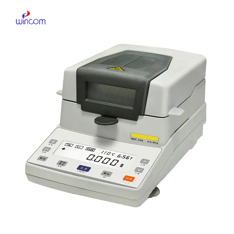
In hospital and research facility settings, digital baby cradling scale provides the critical mass measurement that is needed for delicate analyses. It is the case that reagents, samples, and medicines are weighed with the highest level of precision. Laboratory staff regard the digital baby cradling scale as their helper in making the measurements, carrying out calibrations, and performing quality assurance. Besides being of great assistance in the above activities and ensuring accurate measurement in clinical diagnosis, experimental research, and drug response monitoring, digital baby cradling scale also enhances overall laboratory performance and has a positive impact on the dependability of analytical results.

In pathology laboratories, digital baby cradling scale finds its usage during the staining compounds preparation and the tissue processing additives application. The proper mass measurement guarantees the same composition of the reagent, and this, in turn, affects the performance of the stain and the interpretation under the microscope. This application helps to maintain the same standards in pathology workflows and minimizes the differences between test batches. By using strict preparation conditions, digital baby cradling scale plays a role in the stability of diagnosis in hospital pathology departments.
The era of digital baby cradling scale in hospitals will go beyond the traditional settings and embrace multidisciplinary research environments. As the partnership of clinical, pharmaceutical, and biomedical teams becomes more robust, the analytical balances will cater to different experimental needs. By taking on various analytical actions, digital baby cradling scale will still be a fundamental tool in contemporary hospital laboratory ecosystems.
One of the main tasks in the maintenance of digital baby cradling scale in the hospital laboratory is monitoring the environmental exposure. The presence of excess humidity, direct sunlight, and temperature changes should be completely ruled out. Draft shields should always be kept in a clean and working condition to cause the least possible disturbance in air during the process of weighing. These preventive activities not only help to achieve stable measurements but also aid to lessen the variability in analytical data coming from different medical testing environments.
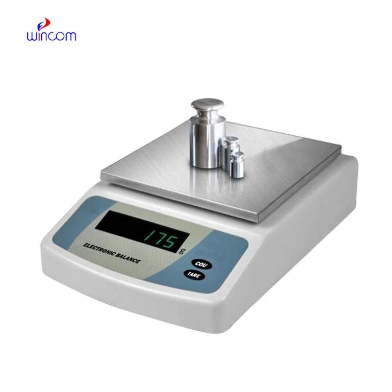
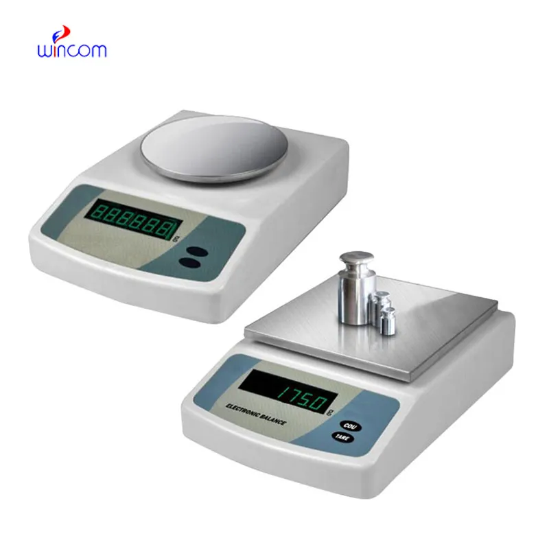
digital baby cradling scale is of great importance for developing and validating laboratory methods. One of the most important prerequisites for getting correct analytical results is the weighing of reference standards, buffers, and chemicals with absolute accuracy. Laboratory personnel depend on digital baby cradling scale for constant concentration reproduction and hence, reliable testing. Its superb sensitivity plus reliable performance indeed make it a primary instrument for validating methods in the clinical, hospital, and pharmaceutical lab settings.
Q: What is the impact of temperature on the performance of analytical balance? A: The changes in temperature can lead to drift and weighing inconsistency. Q: Are analytical balances the only ones used in research laboratories? A: They are very important also for other processes such as sample preparation and improving the accuracy of the experiment. Q: How long does it usually take for an analytical balance to warm up? A: Warm-up times differ from one model to another, but an adequate stabilizing period increases the reliability of the measurement. Q: Is it possible for analytical balances to save weighing data? A: Internal memory or external data transfer are the two ways in which many models can achieve this feature. Q: Would it be necessary to undergo training if one wants to operate an analytical balance? A: Basic laboratory training will be enough to make sure that the balance is being used correctly.
The centrifuge operates quietly and efficiently. It’s compact but surprisingly powerful, making it perfect for daily lab use.
This ultrasound scanner has truly improved our workflow. The image resolution and portability make it a great addition to our clinic.
To protect the privacy of our buyers, only public service email domains like Gmail, Yahoo, and MSN will be displayed. Additionally, only a limited portion of the inquiry content will be shown.
We’re interested in your delivery bed for our maternity department. Please send detailed specifica...
I’m looking to purchase several microscopes for a research lab. Please let me know the price list ...
E-mail: [email protected]
Tel: +86-731-84176622
+86-731-84136655
Address: Rm.1507,Xinsancheng Plaza. No.58, Renmin Road(E),Changsha,Hunan,China

 af
af
 es
es
 ar
ar
 tr
tr
 sw
sw
 pt
pt
 th
th
 ur
ur
 bn
bn
 ne
ne
 vi
vi
 km
km
 lo
lo
 de
de
 ru
ru
 fi
fi
 nl
nl
 fa
fa
 fr
fr
 ko
ko