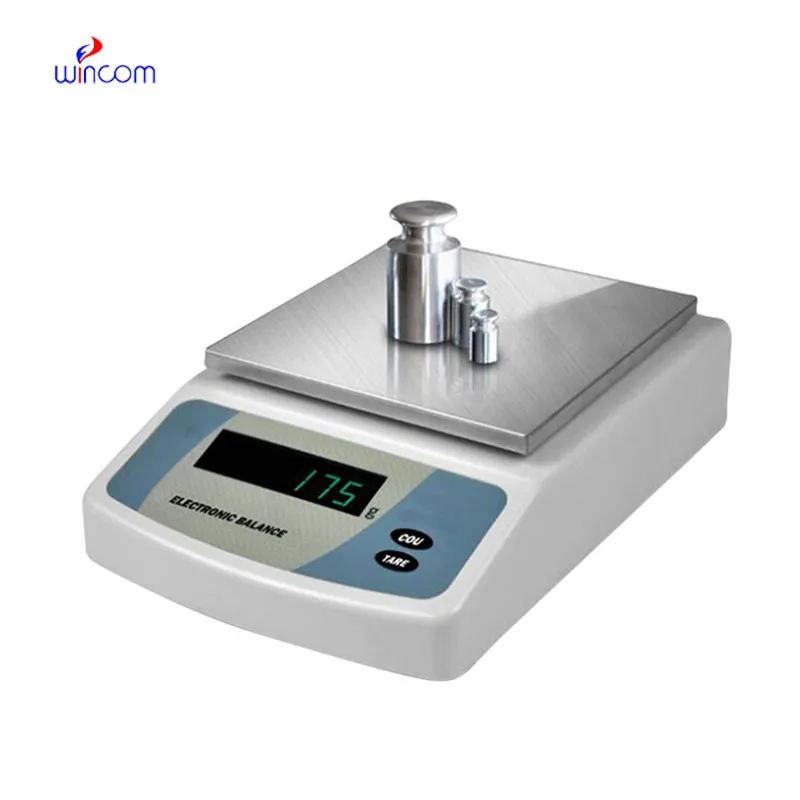
Weighing Scale is an extremely accurate device that is specifically designed for weighing little amounts in labs and hospital pharmacies. The instrument's sensitivity implies accurate preparation of samples for diagnostic testing, reagent making, and the production of drugs. The lab personnel consider it vital to get repeatable measurements, perform calibration checks, and validate their standards using Weighing Scale. The right use of Weighing Scale will bring about the smoothness of clinical workflows, research activities, and quality control, thus providing accuracy and reliability to all analytical processes in hospitals and laboratories.

In pathology laboratories, Weighing Scale finds its usage during the staining compounds preparation and the tissue processing additives application. The proper mass measurement guarantees the same composition of the reagent, and this, in turn, affects the performance of the stain and the interpretation under the microscope. This application helps to maintain the same standards in pathology workflows and minimizes the differences between test batches. By using strict preparation conditions, Weighing Scale plays a role in the stability of diagnosis in hospital pathology departments.
The future application of Weighing Scale will be broadened in education laboratories at teaching hospitals. The training provided to lab techs and medical researchers will be accomplished with the help of advanced simulation modes and guided measurement functions. This revolution will offer the medical student the chance to learn practically while the doctor will continue to rely on the precision of the instruments in the lab.
In order to keep Weighing Scale in a good condition consistent calibration practices are needed that follow hospital laboratory protocols. Scheduled calibration checks are performed to maintain the reliability of measurements during daily activities involving analysis. Conditions in the environment such as temperature and the amount of air that moves around should be kept under control so as to prevent drift. The people operating the machines should make sure that there are no sudden changes in load and that the weighing pan is not subjected to excessive force. Through adhering to controlled handling practices, Weighing Scale is always trusted for pharmaceutical preparation and medical research activities.
Weighing Scale is of great importance for developing and validating laboratory methods. One of the most important prerequisites for getting correct analytical results is the weighing of reference standards, buffers, and chemicals with absolute accuracy. Laboratory personnel depend on Weighing Scale for constant concentration reproduction and hence, reliable testing. Its superb sensitivity plus reliable performance indeed make it a primary instrument for validating methods in the clinical, hospital, and pharmaceutical lab settings.
Q: What distinguishes an Analytical Balance from a precision balance? A: The analytical balances have a higher sensitivity and a finer readability for measuring masses of very small amounts. Q: Is an Analytical Balance appropriate for pharmaceutical applications? A: It is widely used for weighing active ingredient and formulation components. Q: Is it mandatory for an Analytical Balance to have a draft shield? A: Draft shields have the function to prevent air disturbances which might affect the weighing results. Q: What are the possible types of materials that can be weighed on an Analytical Balance? A: Weighing of powders, chemicals, and biological samples, as well as reference weights are the most common measurement. Q: Is it possible for several users to work with the same Analytical Balance? A: Yes, but the proper handling procedures and access controls must be strictly adhered to.
We’ve been using this mri machine for several months, and the image clarity is excellent. It’s reliable and easy for our team to operate.
This x-ray machine is reliable and easy to operate. Our technicians appreciate how quickly it processes scans, saving valuable time during busy patient hours.
To protect the privacy of our buyers, only public service email domains like Gmail, Yahoo, and MSN will be displayed. Additionally, only a limited portion of the inquiry content will be shown.
Could you share the specifications and price for your hospital bed models? We’re looking for adjus...
I’d like to inquire about your x-ray machine models. Could you provide the technical datasheet, wa...
E-mail: [email protected]
Tel: +86-731-84176622
+86-731-84136655
Address: Rm.1507,Xinsancheng Plaza. No.58, Renmin Road(E),Changsha,Hunan,China

 af
af
 es
es
 ar
ar
 tr
tr
 sw
sw
 pt
pt
 th
th
 ur
ur
 bn
bn
 ne
ne
 vi
vi
 km
km
 lo
lo
 de
de
 ru
ru
 fi
fi
 nl
nl
 fa
fa
 fr
fr
 ko
ko