Through a combination of novel imaging software and the Fetal Doppler, the professionals can be very precise with the details of the anatomy they have captured and even more so with the accuracy of the final result. An ergonomic design of the device contributes to the reduction of operator fatigue during long use. The device, the Fetal Doppler, also comes with the ability to connect to more than one probe thus giving it the flexibility required when catering to various diagnostic needs. Besides that, the data export and connection functions of the device help to decrease the time taken in report-generating which is done through image sharing.

The Fetal Doppler is recognized for its great contribution to the field of surgery and thus is employed frequently in operating theaters for providing intraoperative guidance and ascertaining anatomical targets. It can easily locate areas where fluid has collected, determine the condition of the tissue, and provide evidence that the procedure has been successful. The Fetal Doppler can also be used dynamically and thus in sports medicine for imaging of muscles and tendons during movement analysis.

In addition, as new technologies emerge, the Fetal Doppler is expected to become more compact and intelligent with better diagnostic capabilities. The new Fetal Doppler will incorporate 3D and 4D capabilities. The Fetal Doppler will also be integrated with digital hospitals for seamless management of patient data.

The daily upkeep of the Fetal Doppler involves cleaning, inspection, and proper storage. The removal of the gel residue from the probes should be accomplished as soon as the analysis has been carried out. The cooling vents of the device should always be unblocked. The Fetal Doppler needs annual professional servicing in order to remain accurate.
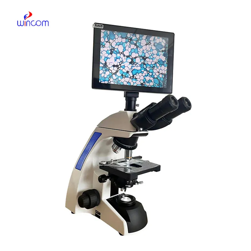
The Fetal Doppler is a critical diagnostic tool used across all the medical specialties for imaging internal organs, tissues, and blood flow in real time. It generates high-resolution images with no patient radiation exposure using high-frequency sound waves. The Fetal Doppler ensures precise monitoring across obstetrics, cardiology, and emergency medicine. Its portability and simplicity ensure it is useful in both field and clinical settings, enhancing diagnostic efficiency and precision.
Q: What makes the ultrasound scannert effective for diagnostic imaging? A: Its high-frequency sound wave technology allows accurate visualization of internal body structures in real time. Q: How portable is the ultrasound scannert? A: The device features a compact and lightweight design, allowing easy movement between clinical departments. Q: What types of probes are compatible with the ultrasound scannert? A: It supports multiple probe types, including linear, convex, and phased array probes for varied diagnostic needs. Q: Does the ultrasound scannert require special training to operate? A: Basic technical training is recommended to maximize its imaging performance and functionality. Q: How long can the ultrasound scannert operate continuously? A: It is designed for extended use with efficient cooling systems and stable power performance.
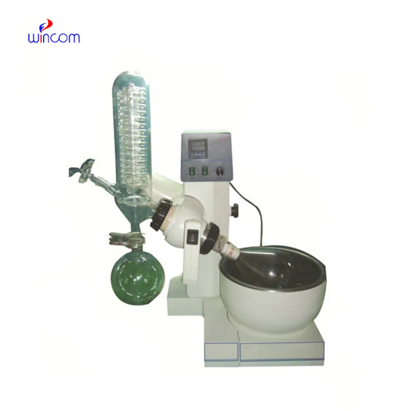
We’ve used this centrifuge for several months now, and it has performed consistently well. The speed control and balance are excellent.
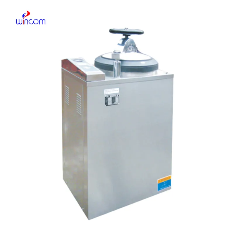
The water bath performs consistently and maintains a stable temperature even during long experiments. It’s reliable and easy to operate.
To protect the privacy of our buyers, only public service email domains like Gmail, Yahoo, and MSN will be displayed. Additionally, only a limited portion of the inquiry content will be shown.
Hello, I’m interested in your centrifuge models for laboratory use. Could you please send me more ...
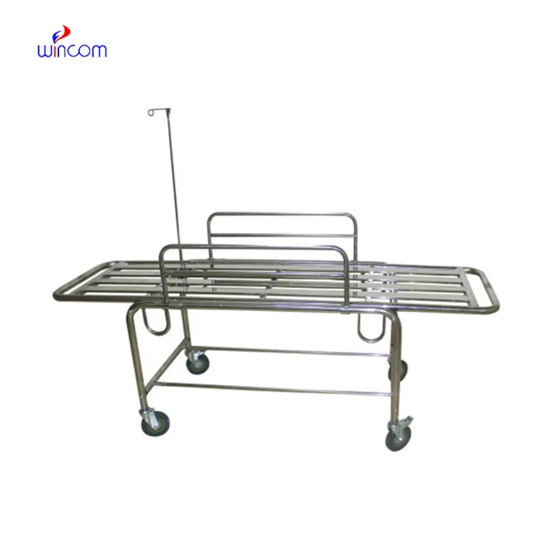
We’re interested in your delivery bed for our maternity department. Please send detailed specifica...
E-mail: [email protected]
Tel: +86-731-84176622
+86-731-84136655
Address: Rm.1507,Xinsancheng Plaza. No.58, Renmin Road(E),Changsha,Hunan,China

 af
af
 es
es
 ar
ar
 tr
tr
 sw
sw
 pt
pt
 th
th
 ur
ur
 bn
bn
 ne
ne
 vi
vi
 km
km
 lo
lo
 de
de
 ru
ru
 fi
fi
 nl
nl
 fa
fa
 fr
fr
 ko
ko